Loculated Pleural Effusion / Loculated Pleural Effusion Radiology Case Radiopaedia Org. Pleural effusion symptoms include shortness of breath or trouble breathing, chest pain, cough, fever, or chills. Pleural fluid/serum protein ratio >0.5. A pleural effusion is accumulation of excessive fluid in the pleural space, the potential space that surrounds each lung. If none is present the fluid is virtually always a transudate. Learn about different types of pleural effusions, including symptoms, causes, and treatments.
ads/bitcoin1.txt
If none is present the fluid is virtually always a transudate. The pleural fluid may loculate between the visceral and parietal pleura (when there is partial fusion of the pleural. Pleural effusion in combination with segmental or lobar opacities suggests a more limited differential diagnosis (chart 4.3). The precise pathophysiology of fluid accumulation varies according to underlying aetiologies. Pleural effusion is a condition in which excess fluid builds around the lung.
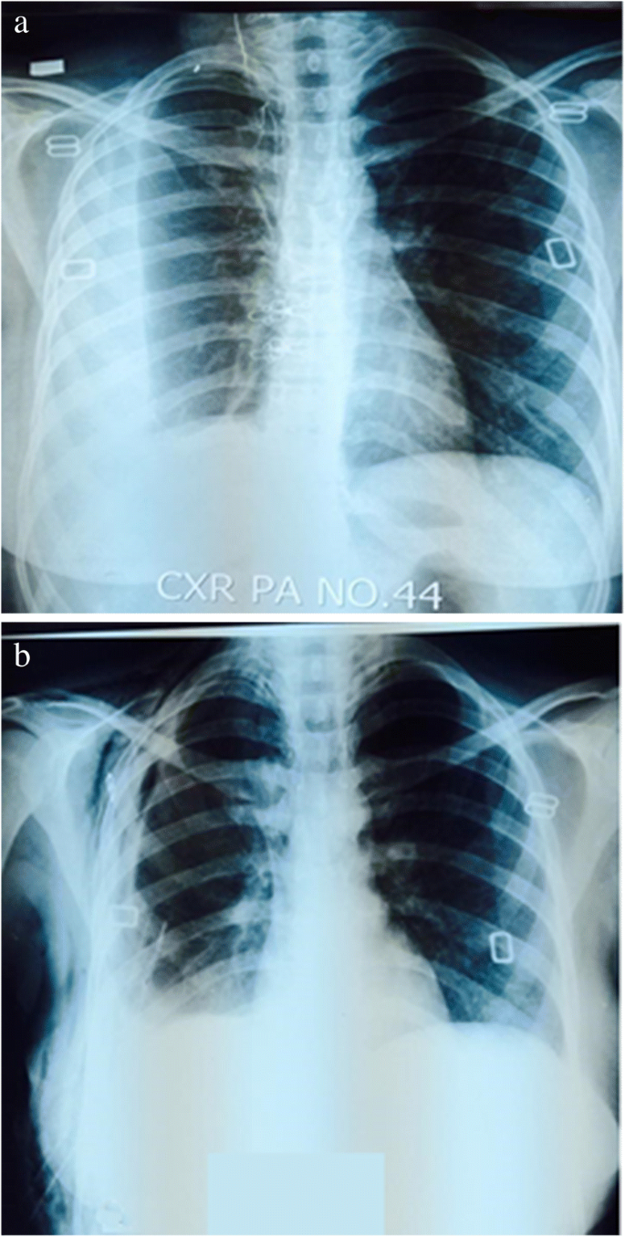
Role Of Medical Thoracoscopy In The Management Of Multiloculated Empyema Bmc Pulmonary Medicine Full Text from media.springernature.com A pleural effusion is accumulation of excessive fluid in the pleural space, the potential space that surrounds each lung. Case contributed by dr prashant mudgal. Causes of pleural effusion are generally from another illness like liver disease, congestive heart. Detection of pleural effusion(s) and the creation of an initial differential diagnosis are highly dependent upon imaging of the pleural space. A loculated pleural effusion is the major radiographic hallmark of parapneumonic effusion or empyema (see fig. Pleural effusion with segmental and lobar opacities. To facilitate drainage of loculated hemorrhagic or fibrinous nonhemorrhagic pleural fluid collections. Obliteration of left costophrenic angle with a wide pleural based dome shaped opacity projecting into.
Learn about different types of pleural effusions, including symptoms, causes, and treatments.
ads/bitcoin2.txt
If none is present the fluid is virtually always a transudate. Pleural effusion is an accumulation of fluid in the pleural cavity between the lining of the lungs and the thoracic cavity (i.e., the visceral and parietal pleurae). The precise pathophysiology of fluid accumulation varies according to underlying aetiologies. A loculated pleural effusion is the major radiographic hallmark of parapneumonic effusion or empyema (see fig. Pleural infection pleural inflammation pleural malignancy (most often pleural fluid analysis findings: Pleural fluid/serum ldh ratio >0.6. Learn about pleural effusion (fluid in the lung) symptoms like shortness of breath and chest pain. A pleural effusion is accumulation of excessive fluid in the pleural space, the potential space that surrounds each lung. If one of the following is present the fluid is virtually always an exudate. Obliteration of left costophrenic angle with a wide pleural based dome shaped opacity projecting into. Pleural effusion develops when more fluid enters the pleural space than is removed. A role in selected clinical circumstances. Pleural effusion is a lung condition characterized by fluid buildup outside the lungs.
In transudative effusion, specific gravity is below 1.015 and. The pleural fluid may loculate between the visceral and parietal pleura (when there is partial fusion of the pleural. Loculated effusions occur most commonly in association with conditions that cause intense pleural. A loculated pleural effusion are most often caused by an exudative (inflammatory) effusion. Pleural fluid/serum protein ratio >0.5.
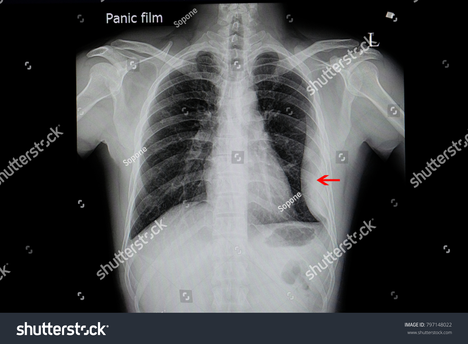
Chest Xray Film Patient Loculated Pleural Stock Photo Edit Now 797148022 from image.shutterstock.com Pleural effusion symptoms include shortness of breath or trouble breathing, chest pain, cough, fever, or chills. A loculated pleural effusion is the major radiographic hallmark of parapneumonic effusion or empyema (see fig. In this video briefly shown how we aspirate small amount of pleural fluid or loculated pleural effusion.for more videos please subscribe the channel.if you. .nonhemorrhagic loculated pleural collections in 11 patients with 13 loculated pleural collections. Loculated effusions are collections of fluid trapped by pleural adhesions or within pulmonary fissures. The precise pathophysiology of fluid accumulation varies according to underlying aetiologies. Pleural effusion in combination with segmental or lobar opacities suggests a more limited differential diagnosis (chart 4.3). The pleura are thin membranes that line the lungs and the.
Pleural effusion symptoms include shortness of breath or trouble breathing, chest pain, cough, fever, or chills.
ads/bitcoin2.txt
In transudative effusion, specific gravity is below 1.015 and. Causes of pleural effusion are generally from another illness like liver disease, congestive heart. Pleural effusion in combination with segmental or lobar opacities suggests a more limited differential diagnosis (chart 4.3). Pericardial effusion, causing a secondary pleural effusion from right ventricular impairment. Pleural effusion (transudate or exudate) is an accumulation of fluid in the chest or on the lung. Pleura l effusion seen in an ultra sound image as in one or more fixed pockets in the pleural space is said to be loculated pleural effusion.in. Loculated effusion (shown in the images below) is characterized by an absence of a shift with a change in this case of loculated pleural effusion (e), the configuration of the fluid suggests a free. Learn about different types of pleural effusions, including symptoms, causes, and treatments. Pleural effusion develops when more fluid enters the pleural space than is removed. A pleural effusion is accumulation of excessive fluid in the pleural space, the potential space that surrounds each lung. Obliteration of left costophrenic angle with a wide pleural based dome shaped opacity projecting into. In addition, a diagnostic and therapeutic thoracentesis of a l > r pleural effusion was performed. Pleural fluid/serum ldh ratio >0.6.
If none is present the fluid is virtually always a transudate. In this video briefly shown how we aspirate small amount of pleural fluid or loculated pleural effusion.for more videos please subscribe the channel.if you. Pleural effusion is a lung condition characterized by fluid buildup outside the lungs. Pleural infection pleural inflammation pleural malignancy (most often pleural fluid analysis findings: The pleura are thin membranes that line the lungs and the.
Role Of Medical Thoracoscopy In The Management Of Multiloculated Empyema Bmc Pulmonary Medicine Full Text from media.springernature.com The precise pathophysiology of fluid accumulation varies according to underlying aetiologies. Pleural infection pleural inflammation pleural malignancy (most often pleural fluid analysis findings: In addition, a diagnostic and therapeutic thoracentesis of a l > r pleural effusion was performed. .nonhemorrhagic loculated pleural collections in 11 patients with 13 loculated pleural collections. If none is present the fluid is virtually always a transudate. Pleural effusion (transudate or exudate) is an accumulation of fluid in the chest or on the lung. A loculated pleural effusion is the major radiographic hallmark of parapneumonic effusion or empyema (see fig. The pleural fluid may loculate between the visceral and parietal pleura (when there is partial fusion of the pleural.
Pleural fluid ldh > two thirds of upper limit for serum ldh.
ads/bitcoin2.txt
Pleural effusion is a condition in which excess fluid builds around the lung. Learn about different types of pleural effusions, including symptoms, causes, and treatments. Case contributed by dr prashant mudgal. .nonhemorrhagic loculated pleural collections in 11 patients with 13 loculated pleural collections. Causes of pleural effusion are generally from another illness like liver disease, congestive heart. In transudative effusion, specific gravity is below 1.015 and. Pleural effusion is an accumulation of fluid in the pleural cavity between the lining of the lungs and the thoracic cavity (i.e., the visceral and parietal pleurae). The precise pathophysiology of fluid accumulation varies according to underlying aetiologies. A role in selected clinical circumstances. Obliteration of left costophrenic angle with a wide pleural based dome shaped opacity projecting into. Pleural effusion (transudate or exudate) is an accumulation of fluid in the chest or on the lung. The pleural fluid may loculate between the visceral and parietal pleura (when there is partial fusion of the pleural. Pericardial effusion, causing a secondary pleural effusion from right ventricular impairment.
ads/bitcoin3.txt
ads/bitcoin4.txt
ads/bitcoin5.txt
0 Response to "Loculated Pleural Effusion / Loculated Pleural Effusion Radiology Case Radiopaedia Org"

0 Response to "Loculated Pleural Effusion / Loculated Pleural Effusion Radiology Case Radiopaedia Org"
Post a Comment